
Rebecca is an equine vet currently helping to teach students and local vets on our Working Equine programme in southern India. On their very first day, Lily Rose was brought to the clinic by her owner and was in need of vital treatment… Rebecca tells us her story!
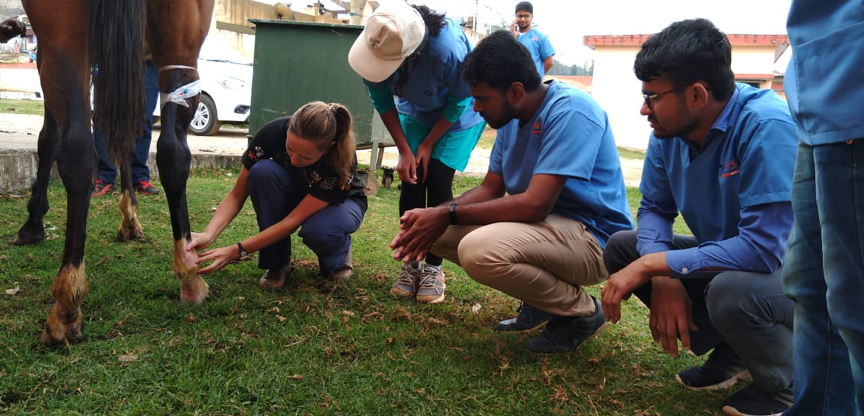
Lily Rose was brought to our clinic on the first day of course in Ooty. She was very lame on her right hind leg and could not place her foot on the ground. Her owner told us that she had stood on a piece of glass a few days previously. He had removed the glass from her frog (the soft triangular part in the underside of her hoof), but he was not sure if he had managed to take out the whole piece.

Lily’s leg was swollen from her foot up to her hock, causing cellulitis (an inflammation of the connective tissues of the leg, causing swelling) and she was in a lot of pain. Penetrating objects to the frog can damage or cause infection in critical structures within the foot, including the coffin bone (small bone within the hoof), deep digital flexor tendon sheath (which helps position and support the foot) and the coffin joint, amongst others. Therefore, aggressive and early treatment is necessary. Our suspicion was that there was either some glass still in her foot, an infection (or hoof abscess) had formed, or that a deeper structure in her body had been damaged. We were working in field conditions at these outreach clinics so could not rely on equipment you would usually have in a clinic; as we could not radiograph the foot, we could only rely on examining the hoof and placing a poultice bandage (specialised moist material which draws foreign material away from a wound) to try to remove any further glass, debris or pus. We treated Lily Rose with broad spectrum antibiotics, anti-inflammatories and pain relief as well as giving her a tetanus injection. We placed a poultice on her hoof and bandaged up to her hock to counteract the swelling of her limb.

To increase the concentration of antibiotic within the foot, we performed intravenous regional perfusion on multiple days. This involves injecting the antibiotic into a vein that is close to the affected area and applying a tourniquet higher up the leg. The tourniquet is left on for 15-20 minutes to temporarily stop the flow of blood to and from the limb, giving the antibiotic more time to perfuse through the tissues. After daily treatment for five days, Lily Rose had shown significant improvement and was walking well without any pain relief!